
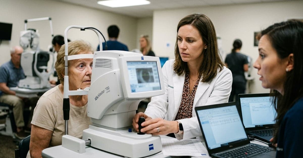
On August 25, 2026, Healio highlighted research indicating that pupil dilation can shift key ocular measurements, potentially skewing IOL power calculations used for cataract surgery planning. The report cites a study in the Journal of Refractive Surgery that found dilation increased anterior chamber depth and reduced lens thickness—two inputs that modern formulas often consider.
What Healio’s report adds for IOL power calculations
The Healio item points to a practical risk: taking biometry after dilation may feed altered anterior segment values into formulas and nudge the refractive target off course. Many contemporary approaches, including Barrett Universal II, incorporate variables beyond axial length and keratometry. They weigh parameters like anterior chamber depth (ACD) and lens thickness (LT), which are sensitive to the eye’s accommodative state. The AAO EyeWiki overview on IOL power calculation lays out how these inputs factor into accuracy across modern formulas.
That’s the crux: if dilation changes ACD and LT, feeding those values into fourth‑generation formulas can alter the selected lens power. According to Healio’s summary of the Journal of Refractive Surgery paper, dilation deepened ACD and thinned the crystalline lens. Those shifts align with long‑observed effects of cycloplegia on accommodation and lens position, discussed in ophthalmic references like EyeWiki’s cycloplegics entry. Even small changes in ACD and LT can tilt a calculation when the formula treats those measures as predictive.
How dilation changes the measurements optometrists rely on
When the ciliary muscle relaxes during pharmacologic dilation, the zonules pull on the crystalline lens. The lens flattens slightly and thins; the anterior chamber can appear deeper. Clinicians have reported these patterns for years in the context of cycloplegic refraction and anterior segment imaging. The Healio-cited study puts that observation into a cataract planning frame: if the biometer records those post-dilation values, the numbers represent the dilated state, not the eye’s typical resting state.
Optical biometers and anterior segment devices are precise, but they are not indifferent to physiology. Consistency of test conditions matters. The EyeWiki page on IOL power calculation emphasizes the role of high‑quality measurements in reducing refractive surprises. Healio’s report adds a practical variable to watch: the timing of drops relative to measurement capture.
Workflow changes to protect IOL power calculations
The signal for optometry practices that comanage cataract patients is straightforward. If dilation can alter ACD and LT, keep measurement conditions steady and predictable. In clinics where comprehensive exams and pre‑op testing happen on the same day, sequence becomes policy, not preference. Several steps can reduce avoidable variance and protect IOL power calculations:
- Capture biometry and anterior segment scans before instilling dilating or cycloplegic drops.
- Document the dilation state in the record when measurements are taken, so surgeons know the context.
- Use the same device and measurement protocol across visits when possible; avoid mixing pre‑ and post‑dilation datasets.
- If a patient returns for repeat measurements, reproduce the original conditions—lighting, fixation, and dilation state.
None of this replaces surgeon preference or local protocol. It sets a default that reduces unexplained spread in the inputs. For practices that share data with surgery centers, a one‑page checklist covering measurement sequence, device, and drop timing can prevent a cascade of small changes that add up to a measurable refractive miss.
Why formula choice and inputs matter as much as timing
Formula selection determines how sensitive the plan is to changes in ACD and LT. Legacy formulas depend mainly on axial length and keratometry. More recent methods, like Barrett Universal II, model effective lens position using a richer set of inputs, including ACD and sometimes LT. As formulas gain predictive power from additional parameters, they also inherit the variability of those parameters. A small, consistent bias in a measurement can map to a consistent, avoidable refractive shift.
Clinicians do not need to abandon dilation on testing days. They need to know which measurements feed which formulas and collect those numbers before drops. The EyeWiki review of IOL power calculation underscores that accurate, reproducible inputs are the bedrock of lens selection. Healio’s pointer to the Journal of Refractive Surgery study simply moves “dilation timing” higher on the list of variables to control.
What to watch next for optometry and surgery teams
Two details will matter as more data emerge: device type and magnitude of effect. Optical biometers differ in how they capture ACD and LT; anterior segment OCT adds its own methods and landmarks. The Journal of Refractive Surgery paper cited by Healio suggests a direction of change under dilation; future reports should clarify how big those shifts are across devices and patient groups. That will help clinics decide when a repeat measurement is warranted.
Communication between optometry and surgery teams is the other lever. Shared protocols for measurement sequence, notation of dilation state, and when to repeat scans can keep a patient on the intended refractive track. For patients with borderline choices between two lens powers, reducing even a small source of variance is worthwhile.
The takeaway is simple and useful: treat dilation timing as part of the measurement protocol, not a footnote. That small change can preserve the integrity of IOL power calculations and cut down on surprises at the slit lamp later. For more on this, see bloomberg.com and nytimes.com.
Related reading: AI Update • Automation • Generative AI